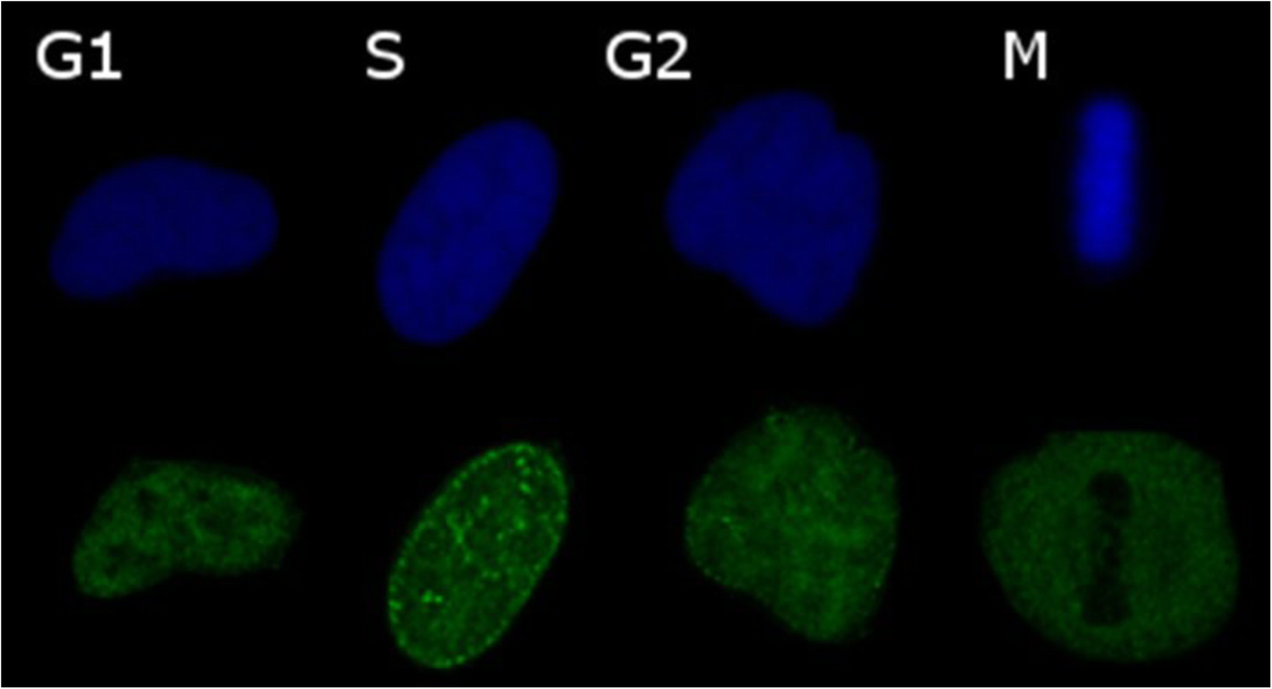
Fig. 1

Confocal laser scanning images of phases of the cell cycle. HeLa nuclei, blue is DAPI green is PCNA. G1 phase is distinguished by solid distribution of PCNA through the nucleus, S phase is distinguished by PCNA speckling through the nucleus and a nuclear border depending on position within S-phase (mid-S displayed here) as well as increased total DNA intensity compared to G1, G2 is distinguished by solid distribution of PCNA and twice the total DNA intensity of G1, M phase is distinguished by the exclusion of PCNA from the nucleus into the cytoplasm